Amsler Grid
The Amsler grid was first developed by Swiss ophthalmologist, Marc Amsler, in 1947. He was a professor of ophthalmology and Chief of the Zurich Eye Clinic at university of Zurich, Switzerland.
The Amsler grid is a one-dimentional square shaped grid with the size of 100mm x 100mm or 10cms x 10cms which is used to measure central 10° field of vision when it is kept at 30cms away from the eye. This grid is portable and provides quick and easy measurements, and this grid can be used to monitor visual distortion at home. Square shaped Amsler Grid is a diagnostic tool used to detect or monitor field of vision in various disorders of macula (macular degenerations, epiretinal membrane), optic nerve and visual pathway due to which metamorphopsia or scotoma can occur. Each square of the grid is 5mm and subtends approximately a 1° angle when kept at 30cms away from the eye.
The original Amsler grid with white lines on black background was developed by Mark Amsler.
The white-on-black (white lines on black background) Amsler charts are more sensitive to macular changes than the black-on-white (black lines on white background) recording sheets. Most frequently used chart is black-on-white in clinics.
Visual distortions are either optical, macular, or cerebral in origin: optical causes include high corneal, lenticular or retinal (staphylomatous) astigmatism, high ametropia, anisometropia, and new glasses.
How to perform:
1. Seat the patient comfortably.
2. While perforning the test room should has good illumination.
3. Test should be performed with best corrected near visual acuity (better to use single vision glass or trial lens). Progressive lenses should be avoided during the test.
4. Keep the Amsler grid at a distance of 30cm away from the eye.
5. Test should be performed with one eye. Better to start test with good eye and occlude other eye properly.
6. Ask the patient to look at the dot located in the center of grid.
7. After fixating at dot located in the center of the grid, the patient should notice all four corners of the grid at a time without moving his/her eye from the dot.
8. If all four corners are not stretched or distorted then the patient should look at the horizontal and vertical lines. If the patient describes the lines as missing or distorted then patient should draw the same on the grid.
In which cases can the Amsler chart be performed?
• Diabetic retinopathy
• Metamorphopsia or visual distortion or wavy vision
• Age related maculopathy
• Hydroxychloroquine (HCQ) user
• Trauma (Berlin's edema)
• Central Cerous Retinopathy (CSR)
There are different types of Amsler Grid to use in different types of retinal eye diseases. So, There are seven different types of Amsler Grid.
Chart 1: This is the basic standard grid which is the more familiar and widely used grid among all the Amsler grids. It consists of a 5 mm square, the white grid on a black card with a central white fixation target, and each square subtending approximately 1° from 30 cm away from the eye. This grid is used to identify various form of distortions as well as Relative and absolute scotoma. Grid with black line on white background is also available.
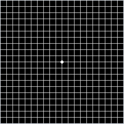
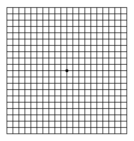
Chart 2: Grid 2 is similar to grid 1 but it consist of two diagonal white lines to assist correct fixation in patients with a central scotoma.
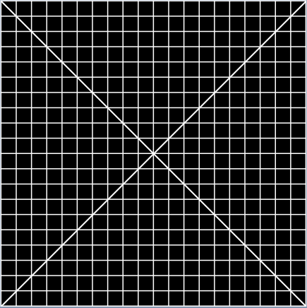
Chart 3: Grid 3 is similar to grid 1 but it has a red grid on a black card with a central red fixation target to stimulate long-wavelength foveal cones. It is used to detect color scotomas and desaturation which may occur in toxic amblyopias, toxic maculopathies, optic neuropathies, and chiasmal lesions but is also capable of testing the malingerer when used with red and green filters.
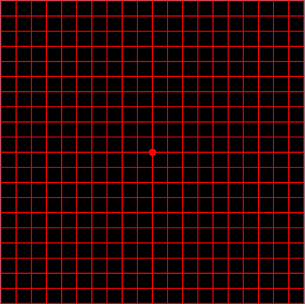
Chart 4: Grid 4 consists of scattered white dots with a central white fixation target on black background. It is used mainly to distinguish scotoma from metamorphopsia, as there is no form to be distorted.
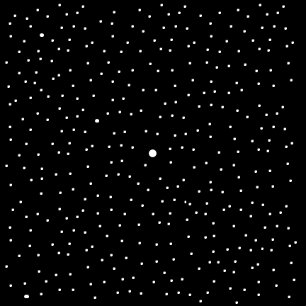
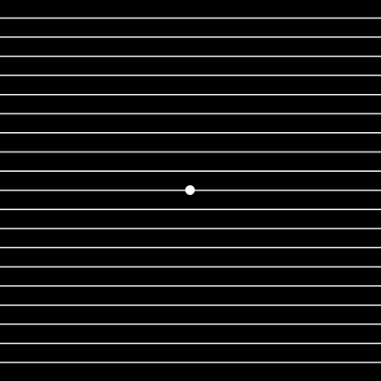
Chart 6: Grid 6 is similar to grid 5 but it consist of black lines on a white card with additional lines at 0.5° above and below fixation. With this grid metamorphopsia along with the reading, the level can be easily detected.
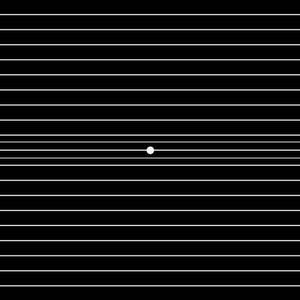
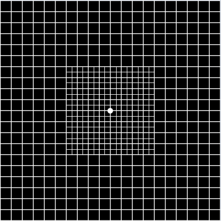
Chart 7: Grid 7 is similar to grid 1 but with additional 0.5° squares in the central 4°. Grid 7 exhibits a fine central grid and is used for the detection of subtle visual disturbance from macular disease, especially in early disease.